 Biochemistry
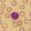 Haematology and Transfusion
 Clinical Microbiology (Including Virology)
 Cellular Pathology
 General Information
 Immunology
 Molecular Pathology
![]()
Preferred Sample Type
")
Suitable Specimen Types
SerumLactate Dehydrogenase (LDH)
Specimen Volume
1mL blood (250ul serum)Sample Preparation
Centrifuge specimen. Serum samples in non-gel tubes should be separated from cells promptly.
Turnaround Time
1 daySample Processing In Laboratory
Usual
Sample Stability
4 days at 2 ºC to 8 ºCGeneral Information
Lactate dehydrogenase is a widespread cytosolic enzyme, found in the greatest concentrations in heart, skeletal muscle, liver, kidney and red blood cells. Many of these tissues rely on anaerobic glycolysis for energy. LDH catalyses the conversion of pyruvate to lactate, and vice versa. In the forward reaction, NADH is oxidized to produce NAD. NAD is vital as an oxidizing agent to facilitate flux through the glycolytic pathway. Increased plasma activity is usually the result of leakage of enzyme from tissues as a result of damage: it is 500 times more concentrated within the cytosol relative to plasma. LDH is a tetramer made up of combinations of 2 polypeptide chains. Five isoenzymes are recognised, designated LD1 to LD5, according to the number of each chain type resulting in different molecular mass and electrophoretic mobility:
- LD1 and LD2 are predominant in the heart, red cells and kidney
- LD4 and LD5 are predominant in the liver and some skeletal muscles
- No single isoenzymes predominate in the lung and spleen
- LDH may rise following myocardial infarction and in myocarditis.
Following a myocardial infarction:
- levels increase after 12 to 24 hours
- peak at 48 to 72 hours
- return to normal after 8 to 10 days, providing there are no complications
- the peak reached is from two to ten times the upper reference limit
- the normal plasma LD1:LD2 isoenzyme ratio of less than 1.0 is reversed
Therefore, LDH was thought to provide a valuable late indicator of infarction. It is however not often used in this context in current practice.
LDH may also be raised in:
- liver disease - poor specificity
- skeletal muscle disease e.g. muscular dystrophies
- haemolysis, both in vitro and in vivo • leukaemia - to very high levels
- pernicious anaemia - very high levels
- megaloblastic anaemia - very large elevation
- myeloproliferative disorders
- thalassaemia
- malignancy of all types e.g. raised levels in pleural fluid relative to plasma are a key index of suspicion
- renal infarction
- pulmonary embolus
- congestive cardiac failure
- hypothyroidism
Patient Preparation
None
Notes
Analysis CANNOT be performed on specimens that have been refrigerated or frozen.
Haemolysis interferes with this method
Please note Isoenzyme analysis is not available
Reference Range
125 - 220 U/L
Source of Reference Range
Abbott DiagnosticsSpecifications
- EQA Status: NEQAS
- EQAS Scheme: Yes